Model species for marine biotechnology
Per definition, model species are organisms that are chosen by scientists for in-depth studies. They are normally chosen in function of a certain property of the organism, or their potential to provide answers to universal questions. They are handy because the information gained about these organisms is transferable to other living organisms on which it is unfeasible to perform in-depth studies. Model organisms have to meet certain criteria such as a short generation time, the possibility to cultivate them in the lab, low costs associated, the possibility to easily genetically manipulate, having a feasible genome size, being easy to work with and having an interesting evolutionary position.
Terrestrial model species can also provide some answers to marine biotechnological questions, but the focus here is on marine model species, although there are not as many model species from the marine environment than from the terrestrial environment. Generally a model organism is not chosen in function of biotechnological research but for fundamental biology research. This does not mean that the discoveries coming from this type of research do not provide interesting information about biotechnological applications. An overview of marine model organisms is available[1], but there is a need for an in-depth evaluation to identify, prioritise and select a limited number of appropriate marine model organisms which could provide critical knowledge to stimulate the development of biotechnological applications. Below, some marine model species which are very useful for biotechnological applications are being discussed.
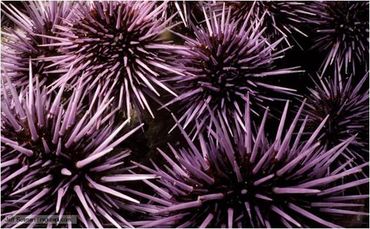
The California Purple Sea Urchin, Strongylocentrotus purpuratus, is a good model for cancer research or neurodegenerative disorders.[2] Sea urchins in general provide a unique opportunity to study patterns in developmental biology and cell cycle regulation, as this group is evolutionary more closely related to humans than other major invertebrate models in use. Almost everything we know about the chromosomal basis of development, maternal determinants, fertilization and maternal messenger RNA originates from proper sea urchin research. The knowledge generated using sea urchin studies thus gives the opportunity to discover potential new targets for cancer therapy in humans.

Fig 1. Strongylocentrotus purpuratus.[3]
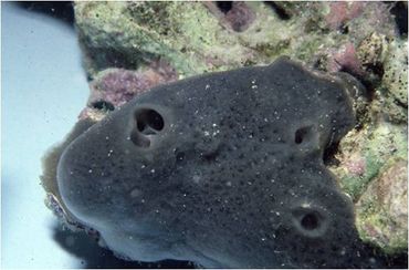
The demosponge Amphimedon queenslandica, native to the Great Barrier Reef[4], is another marine model species on which research is conducted in function of evolutionary developmental biology and comparative genomics.[5] Research conducted on these organisms is interesting for biotechnological applications because the sponge typically hosts large and diverse communities of microbes[6] or has a symbiotic relationship with these microbes. These microbes may be a potential source of secondary metabolites.[7] The examination of the relationship between host-microbes also allows us to learn more about the metabolic pathways in these ‘primitive’ organisms.[8]

Fig 2. Amphimedon queenslandica.[9]
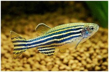
Zebrafish (Danio rerio) are used for different applications. One of the main reasons zebra fish have become model organisms is because of their transparency during development, allowing us to study the processes taking place inside the fish. These organisms have already provided great advancements in the field of vertebrate development and disease modeling. It is used to model infectious diseases that affect humans.[10] They are studied to find solutions for the widespread problems of diseases found in wild and farmed fish.[11] Another important feature of zebrafish worth mentioning is the possibility to become colonized by aquatic pathogens without causing disease. This makes them excellent models for the study of fish acting as a pathogen reservoir.[12]

Fig 3. Danio rerio.[13]
Many more marine organisms are nowadays investigated in Europe. This is interesting for biotechnological studies because of the constant discovery of new applications and molecules, such as:
- New enzymes
- Proteins and peptides
- Secondary metabolites
- Polysaccharides (bacteria, archaea, algae, marine plants)
- Fatty acids and lipids (microalgae).
References
- ↑ Srivastava, M. (2010).The Amphimedon queenslandica genome and the evolution of animal complexity. Nature. Vol. 466: 720-727
- ↑ Braun, T; Woollard, A; RUNX factors in development: Lessons from invertebrate model systems (2009); Volume: 43; Issue: 1; Pages: 43-48
- ↑ http://www.bbc.co.uk/nature/life/Strongylocentrotus_purpuratus
- ↑ Watson, J. R. et al.(2014). Analysis of the Biomass Composition of the Demosponge Amhimedon queenslandica on Heron Island Reef, Australia
- ↑ Srivastava, M. (2010).The Amphimedon queenslandica genome and the evolution of animal complexity. Nature. Vol. 466: 720-727.
- ↑ Thomas, T. R. A., Kavlekar, D. P. and LokaBharathi, P. A. (2010). Marine drugs from Sponge-Microbe Association-A review. Mar. Drugs. 8: 1417-1468
- ↑ Wilson, M. C. et al. (2014). An environmental bacterial taxon with a large and distinct metabolic repertoire. Nature 506: 58-62.
- ↑ name="radax">Radax, R. Hoffmann, F., Rapp, H. T., Leininger, S., and Schleper, C. (2011). Ammonia-oxidizing archaea as main drivers of nitrification in cold-water sponges. Environ. Microbiol. 14: 909-923.
- ↑ name="marinesp">http://www.marinespecies.org/
- ↑ name="davis">Davis, J. M. (2002). Real-time visualization of Mycobacterium-Macrophage interactions leading to initiation of Granuloma formation in zebrafish embryos. Immunity. Vol. 17: 693-702
- ↑ name="neely">Neely, M. N., Pfeifer, J. D. and Caparon, M. (2002). Streptococcus-Zebrafish model of bacterial pathogenesis. Infection and Immunity. Vol. 70 (7): 3904-3914.
- ↑ name="runft">Runft, D. L. et al. (2014). Zebrafish as a natural host model for Vibrio chloreae colonization and transmission. Applied and Environmental Microbiology. Vol 80 (5): 1710-1717.
- ↑ name="clup">http://apps.acesag.auburn.edu/mediamax/pictures/51/clupeiformes-clupeidae.html